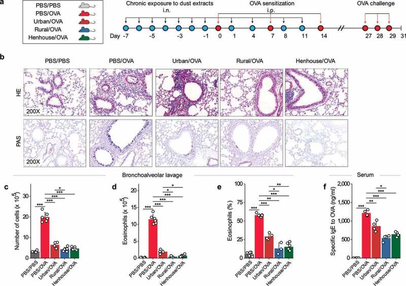
Figure 5.

Repeated nasal inhalation (n = 11) of dust extracts regulated OVA-induced allergic airway inflammation in mice. (a) Study protocol illustrating the dust extracts, intranasal exposure, and sensitization, as well as challenge by OVA. The dust extract corresponds to the mixture of extracts from each group in Figure 3 (solid dot). (b) Histopathology of lung tissue (Hematoxylin and Eosin (H&E) and Periodic Acid-Schiff (PAS) staining) in each group as indicated above. (c–f) Inflammatory cell counts, number and percentage of eosinophils in bronchoalveolar lavage fluid, and serum-specific IgE level against OVA in the serum. * p < 0.05, ** p < 0.01, and *** p < 0.001 indicate the levels of different significant differences calculated by a t test or Wilcoxon test.
