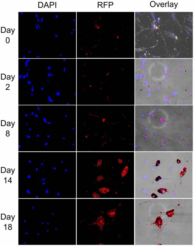
Figure 6.

Oil red O lipid and DAPI staining of SGBS cells at different stages of differentiation from day 0–18 (10× magnification).
Legend: DAPI staining of nuclei in blue, RFP staining of cells in red

Oil red O lipid and DAPI staining of SGBS cells at different stages of differentiation from day 0–18 (10× magnification).
Legend: DAPI staining of nuclei in blue, RFP staining of cells in red