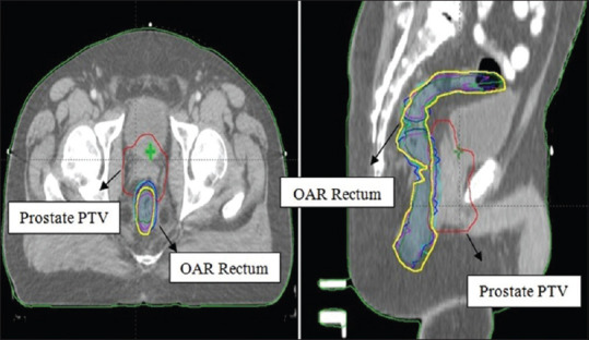
Figure 3.

Superimposed rectal contours of a typical patient on axial (left) and sagittal (right) views as ----- Original rectum volume (yellow contour) in planning CT; ----- Rectum volume in CBCT1; ----- Rectum volume in CBCT2; ----- Rectum volume in CBCT3; ----- Rectum volume in CBCT4; ----- Rectum volume in CBCT5
