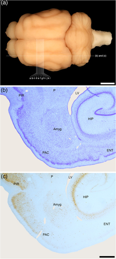
FIGURE 1.

(a) Photograph of the dorsal surface of the tree pangolin brain showing the approximate sagittal plane of the sections depicted in (b) and (c) of this figure, and the approximate coronal planes of the Diagrams (a)–(l) of Figure 2. Scale bar in (a) = 5 mm. Photomicrographs of sagittal (b) Nissl‐stained and (c) parvalbumin immunostained sections through the amygdaloid body (Amyg) of the tree pangolin showing the topological relationships of this structure within the brain of this species. In all images, rostral is to the left, and in (b) and (c) dorsal to the top of the image. Scale bar in (c) = 1 mm and applies to (b) and (c). See list for abbreviations
