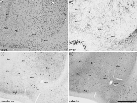
FIGURE 4.

Low magnification photomicrographs of the accessory basal amygdaloid nucleus (AB) within the amygdaloid body of the tree pangolin stained for neuronal nuclear marker, NeuN (a), myelin (b), parvalbumin (c), and calbindin (d). Note that only subtle variations exist between the parts of this nucleus, the magnocellular (ABmc), intermediate (ABi), parvocellular (ABpc) and shell (ABsh) parts, with the cytoarchitecture as revealed with NeuN (a) immunostaining being the most reliable stain for identification of these parts. In all images, dorsal is to the top and medial to the left. Scale bar in (d) = 1 mm and applies to all. See list for abbreviations
