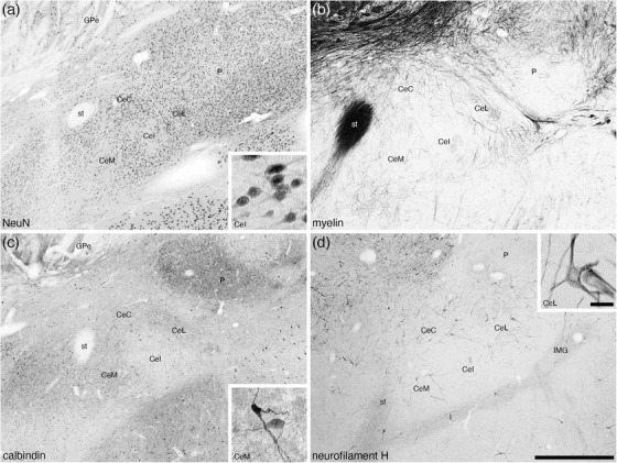
FIGURE 7.

Low magnification photomicrographs of the central nuclear cluster within the amygdaloid body of the tree pangolin stained for neuronal nuclear marker (NeuN, a), myelin (b), calbindin (c), and neurofilament H (d). Although the architectonic delineation into distinct subnuclei is not always unambiguous, we could identify with reasonable certainty the medial (CeM), intermediate (CeI), lateral (CeL), and capsular (CeC) divisions of the central nuclear cluster of the centromedial group. In all images, dorsal is to the top and medial to the left. Scale bar in (d) = 1 mm and applies to all. Scale bar inset (d) = 25 μm and applies to all insets. See list for abbreviations
