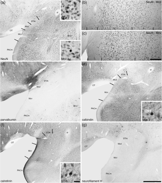
FIGURE 8.

Low and higher magnification photomicrographs of the medial amygdaloid nucleus within the amygdaloid body of the tree pangolin stained for neuronal nuclear marker (NeuN, a, b, c), parvalbumin (d), calbindin (e), calretinin (f), and neurofilament H (g). We observed both dorsal (Mcd) and ventral (Mcv) subdivisions of the medial amygdaloid nucleus, as well as the intramygdaloid division of the bed nucleus of the stria terminalis (STIA). Both Mcd and Mcv evinced three layer (1, 2, 3) (b, c), although the laminar borders were not sharp. The insets in (a) are high magnification images of NeuN immunostained neurons in layer 2 of the Mcd and Mcv, the inset in (e) is of calbindin immunostained neurons in layer 2 of the Mcv, and the inset in (f) is of calretinin immunostained neurons in the STIA. In all images, dorsal is to the top and medial to the left. Scale bar in (g) = 1 mm and applies to (a), (d)–(g). Scale bar in (c) = 200 μm and applies to (b) and (c). Scale bar in inset (f) = 25 μm and applies to all insets. See list for abbreviations
