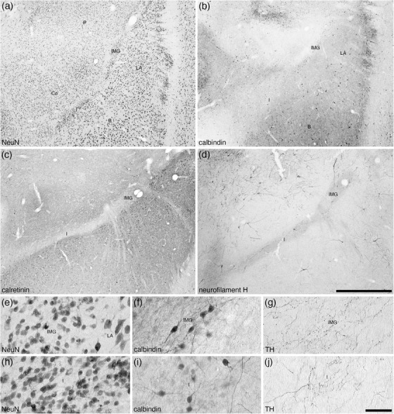
FIGURE 10.

Lower (a–d) and higher (e–j) magnification photomicrographs of the amygdaloid intramedullary gray matter (IMG) and intercalated island, or band, of the amygdala (I) within the amygdaloid body of the tree pangolin stained for neuronal nuclear marker (NeuN, a, e, h), calbindin (b, f, i), calretinin (c), neurofilament H (d), and tyrosine hydroxylase (g, j). Note how both the IMG and I form distinct bands of small neurons (e, h) between the lateral amygdaloid nucleus and putamen (IMG) and basal amygdaloid nucleus and central amygdaloid nucleus (I). Note the specific absence of calretinin immunostaining in these nuclei (c), and the marked neuropil staining for neurofilament H in both nuclei (d). Some of these neurons are immunopositive for calbindin (f, i), and in both the IMG and I there is a moderately dense tyrosine hydroxylase immunopositive terminal network (g, j). In all images, dorsal is to the top and medial to the left. Scale bar in (d) = 1 mm and applies to (a)–(d). Scale bar in (j) = 50 μm and applies to (e)–(j). See list for abbreviations
