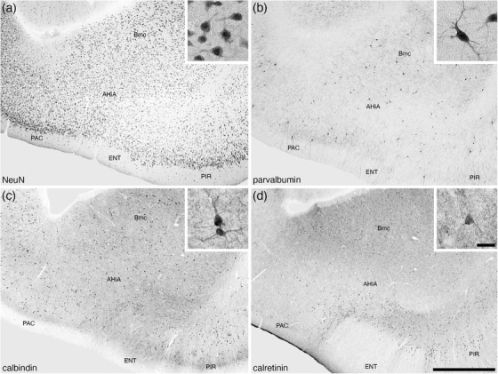
FIGURE 11.

Lower (a–d) and higher (insets of a–d) magnification photomicrographs of the amygdalohippocampal area (AHiA) of the amygdaloid body of the tree pangolin stained for neuronal nuclear marker (NeuN, a), parvalbumin (b), calbindin (c), and calretinin (d). The insets are high magnification images of immunostained neurons in the CeI (NeuN, inset a), CeM (calbindin, inset c), and CeL (neurofilament H, inset d). In all images, dorsal is to the top and medial to the left. Scale bar in (d) = 1 mm and applies to (a)–(d). Scale bar in inset (d) = 25 μm and applies to all insets. See list for abbreviations.
