
FIGURE 2.
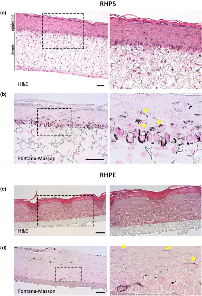
Histology of reconstructed human pigmented skin and epidermis models. Reconstructed human pigmented skin (RHPS) (a) and (b), reconstructed human pigmented epidermis (RHPE) (c) and (d). Haematoxylin and eosin (H&E) staining is shown in (a) and (c), and Fontana‐Masson staining is shown in (b) and (d) to identify pigment in melanocytes and keratinocytes. Dermis is visible in the skin model and the stratification and differentiation of epidermis are visible in both models. Magnified views of the boxed areas are shown in the right panels. Images are representative of three independent experiments. Arrowheads indicate areas of pigment accumulation in keratinocytes. Scale bars, 50 μm