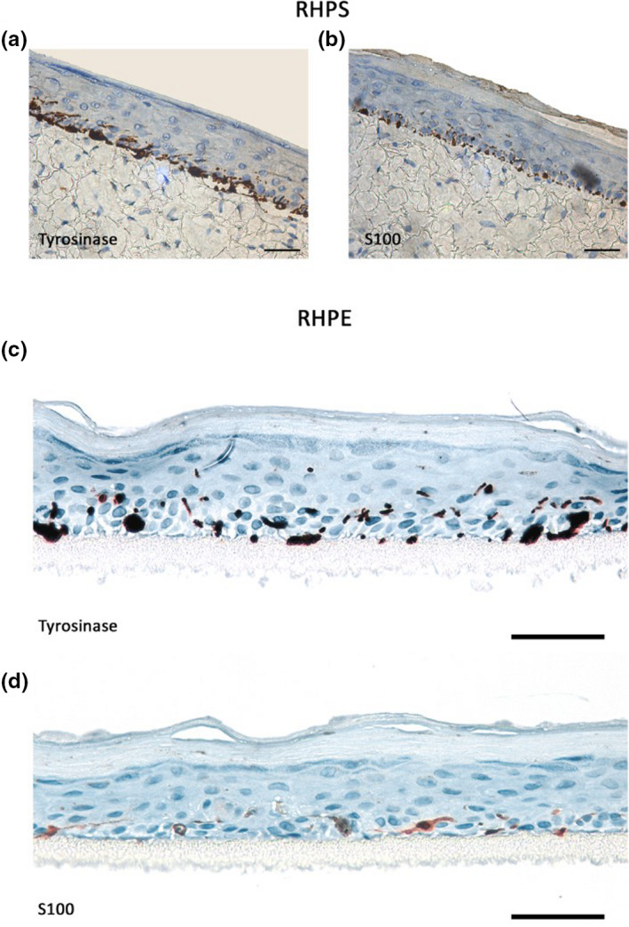
FIGURE 3.

Immunohistochemical analysis of epidermal melanocytes in reconstructed human pigmented skin (RHPS) and reconstructed human pigmented epidermis (RHPE) models. Melanocytes in the basal layer were identified by staining for tyrosinase or S‐100 proteins in RHPS (a) and (b) and RHPE (c) and (d). Images are representative of three independent experiments. Scale bars, 50 μm
