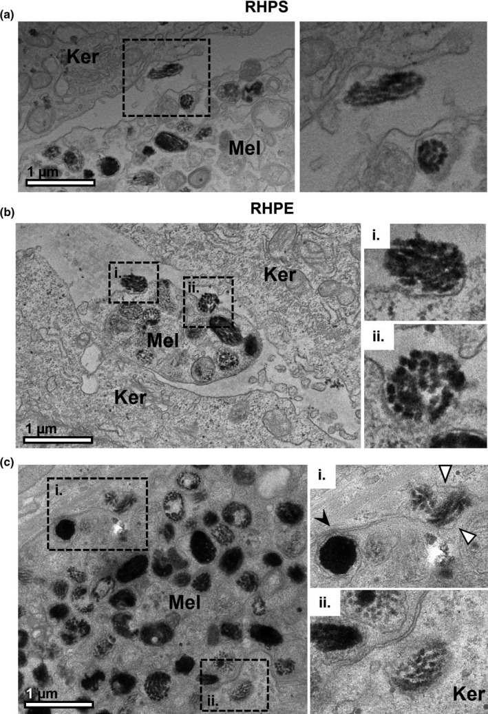
FIGURE 5.

Melanocores in the extracellular space between melanocyte dendrites and keratinocytes. Melanocyte dendrites (Mel) are observed between keratinocytes (Ker) in reconstructed human pigmented skin (RHPS) (a) and reconstructed human pigmented epidermis (RHPE) (b) and (c) by transmission electron microscopy. The boxed regions are shown at higher magnification in the right panel. Several examples of melanocores between the cells and close to melanocyte dendrites are shown in (a), (b) and (c). Melanocores recently exocytosed by the melanocyte are shown in (b) i and (b) ii. A melanosome in the process of fusing with the melanocyte plasma membrane is visible in (c)i., indicated by the black arrowhead, and a melanocore is observed between the melanocyte and keratinocyte plasma membranes, indicated by the white arrowheads. In (c)ii., a recently transferred melanocore is observed in the keratinocyte. Scale bar, 1 μm
