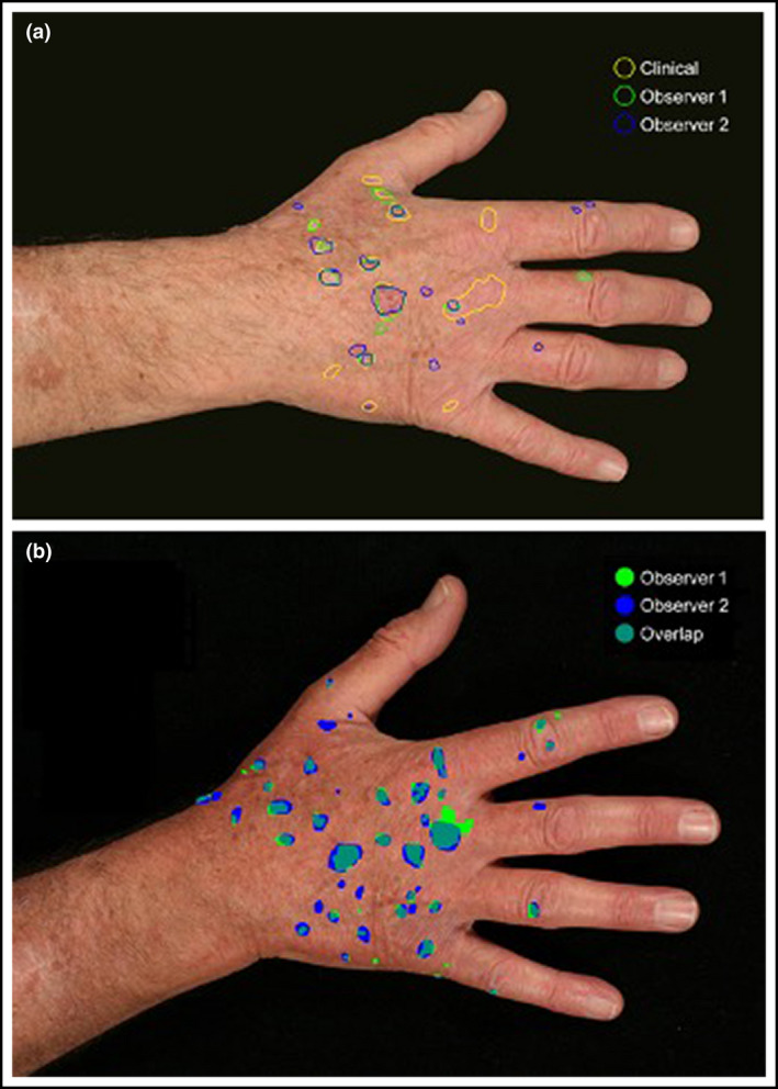
Figure 2.

Digital photographs corresponding with clinical actinic keratosis (AK) assessments were taken at all timepoints. An electronic image capture programme was used by two independent observers (observer 1 and 2) to annotate images. (a) These annotated images were matched with the original clinical assessment (clinical) by two separate observers. These results were used to calculate the sensitivity and false discovery rates for photography vs. clinical assessment of individual AKs. (b) The annotated images were also used to derive a Dice coefficient for interobserver concordance (overlap) in the photographic assessment of AKs annotated by each observer. [Colour figure can be viewed at wileyonlinelibrary.com]
