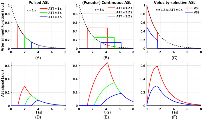
FIGURE 2.

The arterial input function of (A) pulsed arterial spin labeling (PASL) with the temporal width of the labeling bolus of 1.0 s, arterial transit time (ATT) of 1.0, 2.0, 3.0 s; (B) pseudo‐continuous ASL (PCASL) with the labeling duration of 3.0 s, ATT of 1.2, 2.2, and 3.2 s; and (C) velocity‐selective ASL (VSASL) with VS saturation (VSS) and VS inversion (VSI) pulses, and ATT of 0 s, the temporal width of the labeling bolus of 1.6 s. In this illustration of ideal conditions, t is the time from the start of the labeling pulse, the labeling efficiency (for VSS, ), , T1 = 1.6 s. Their corresponding kinetic curves indicate that the maximal perfusion‐weighted signal of pulsed ASL (D) is only half of that of PCASL (E), and both are sensitive to ATT. In contrast, the ATT for VSASL (F) is essentially 0 and the maximal signal of VSI is about (or more than) twice that of PCASL when the ATT of PCASL is longer than 2.2 s. This figure demonstrates that, with its full potential reached, VSASL has minimal susceptibility to ATT effects among existing methods and has a significant signal advantage especially when long ATTs with PASL or PCASL are expected
