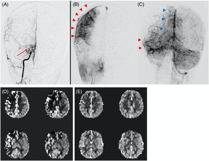
FIGURE 7.

A 6‐year‐old with right‐sided moyamoya status post successful revascularization (Matsushima A). (A) Cerebral angiogram during a right ICA injection demonstrates high‐grade stenosis of the right ICA terminus, MCA origin, and ACA origin (red arrow), with nearly absent antegrade filling/ perfusion of the distal brain parenchyma. (B) Most of the right MCA territory (red arrowheads) is perfused through surgical collaterals via the ECA. (C) Remainder of the MCA (red arrowheads) and ACA (blue arrowheads) territories are perfused by native collaterals via the posterior circulation (left vertebral artery injection). (D) Traditional pulsed arterial spin labeling (PASL) fails to capture this perfusion because of prolonged arterial transit time (ATT)s through the collateral pathways; artifactual perfusion deficits and macrovascular signals are seen. (E) Velocity selective ASL (VSASL) accurately captures perfusion in these territories and is concordant with the angiogram
