Fig. 5.

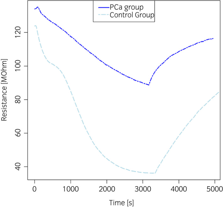
Typical response of one sensor of the eNose array recorded during the analysis of urine headspaces from control subjects (light blue line) and PCa patients (blue line). [Colour figure can be viewed at wileyonlinelibrary.com]

Typical response of one sensor of the eNose array recorded during the analysis of urine headspaces from control subjects (light blue line) and PCa patients (blue line). [Colour figure can be viewed at wileyonlinelibrary.com]