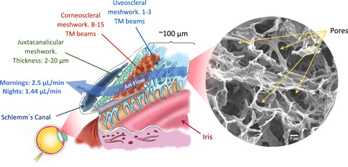
Figure 1.

Location of the trabecular meshwork (TM). The anatomical structure of this tissue formed by three layers: uveal, corneoscleral, and juxtacanalicular meshwork. Scanning electron microscopy (SEM) image of a real decellularized TM tissue (author's own work). Scale bar: 2μm.
