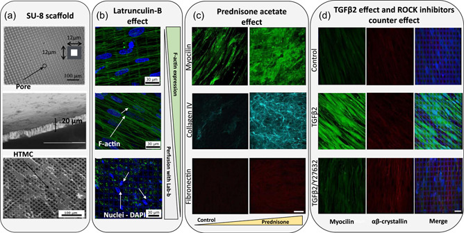
Figure 3.

Different studies that use a SU‐8 scaffold as a TM model. (a) Scanning electron microscopy (SEM) images of the microfabricated SU‐8 scaffold. From top to bottom: Pore size of the scaffold, the cross section and SEM micrographs of human TM cells grown in SU‐8 scaffold with a pore size of 12 μm. Reproduced with permission from Torrejon et al. (2013); John Wiley and Sons. (b) Biological response to 2 μM Latrunculin‐B. Confocal images of F‐actin cytoskeleton in green and co‐stained nuclei with DAPI in blue. From top to bottom: before perfusion, after perfusion with medium, and after perfusion with medium and Lat‐B. Reproduced with permission from Torrejon et al. (2013); John Wiley and Sons. (c) Confocal images of myocilin (green), collagen IV (cyan), and fibronectin (red) after perfusion with 300 nM prednisone acetate. Scale bar: 40 μm. Reproduced with permission from Torrejon, Papke, Halman, Bergkvist, et al. (2016), Torrejon, Papke, Halman, Stolwijk, et al. (2016); John Wiley and Sons. (d) Confocal images of human TM protein expression after treatment with 2.5 ng/ml TGFβ2 in the absence or presence of 10 μM Y27632. From left to right, myocilin in green, αβ‐crystallin in red, and merged images. Scale bar = 30 μm. Reprinted from Torrejon, Papke, Halman, Bergkvist, et al. (2016), Torrejon, Papke, Halman, Stolwijk, et al. (2016) (https://www.nature.com/articles/srep38319).
