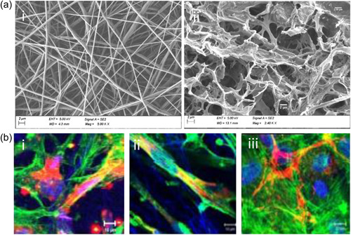
Figure 4.

Electrospinning‐based scaffolds for TM tissue. (a) SEM images showing similarities between: (i) PCL electrospinning‐based scaffold and (ii) native human TM tissue. (Scale bar: 2 µm) (B) Laser scanning confocal microscopy images of TM cells over nanofibers (i) non‐aligned PCL, (ii) aligned PEUU, and (iii) glass, respectively. Nuclei of TM cells in blue (DRAQ 5TM), actin filaments in green (Alexa Fluor 488 phalloidin®), and the ECM materials in red (Alexa fluor 555®). PEUU, poly‐etherurethane urea; SEM, scanning electron microscopy; TM, trabecular meshwork. Reprinted from Kim et al. (2009). Thirteenth International Conference on Miniaturized Systems for Chemistry and Life Sciences.
