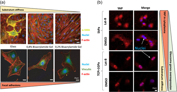
Figure 6.

Substratum stiffness effects. (a) Substrate rigidity modulates α‐SMA localization. HTM cells grown on collagen‐coated coverslips and collagen‐coated stiff (0.8% bis‐acrylamide) or (0.2% bis‐acrylamide) polyacrylamide gels for 10 days. Composite images depict F‐actin in red, α‐SMA in green, and cell nuclei in blue. Some cells exhibit α‐SMA positive stress fibers on stiff polyacrylamide gels, whereas intense staining is observed on glass coverslips. Scale bar: 40 μm. Bottommost images indicate higher focal adhesions on stiffer substrates. F‐actin (red), vinculin (green), and nucleus (blue). Scale bar: 20 μm. Reproduced with permission from Schlunck et al. (2008) Association for Research in Vision & Ophthalmology (ARVO). (B) Substratum stiffness and Lab‐B alters nuclear/cytoplasmic localization in HTM cells. HTMC stained for YAP (red) and counterstained with DAPI (blue). YAP localization in HTMC is mixed between nuclear and cytoplasmic. Nuclear localization is more pronounced on TCP (>1 GPa) than on the 5 kPa hydrogel. With Lat‐B treatment, nuclear YAP localization is decreased on the 5 kPa hydrogels but increased on TCP, which induces higher probabilities to overexpress glaucomatous genes. Scale bar: 10 μm. Reproduced with permission from Thomasy et al. (2013). Elsevier.
