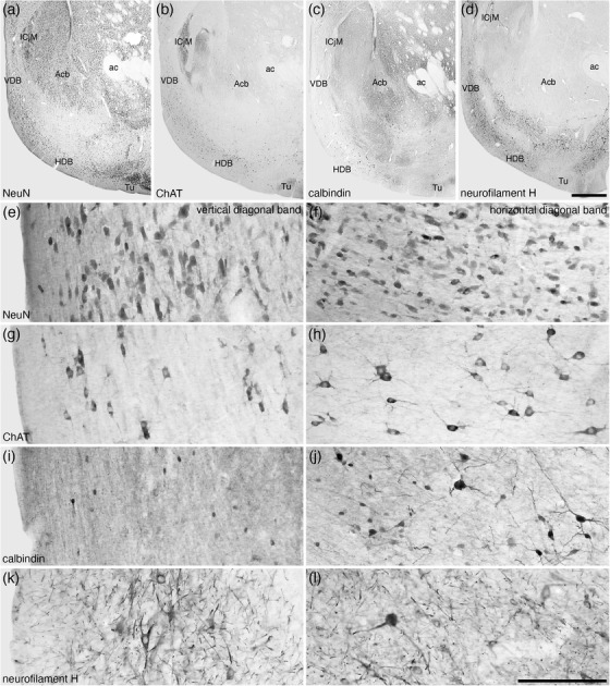
FIGURE 4.

Lower (a–d) and higher (e–l) magnification photomicrographs of the nucleus of the diagonal band (of Broca) in coronal sections of the tree pangolin brain stained for neuronal nuclear marker (NeuN; a, e, and f), choline acetyltransferase (ChAT; b, g, and h), calbindin (c, i, and j), and neurofilament H (d, k, and l). Note the presence of numerous cholinergic neurons in both the vertical diagonal band (VDB, Ch2; g) and horizontal diagonal band (HDB, Ch3; h), larger and smaller calbindin immunopositive neurons in the HDB (j) compared with only smaller neurons in the VDB (i), and the numerous structures immunopositive for neurofilament H in both the VDB and HDB (k and l). In all images, medial is to the left and dorsal to the top. Scale bar in (d) = 1 mm and applies to (a)–(d). Scale bar in (l) = 200 μm and applies to (e)–(l). See list for abbreviations
