
FIGURE 5.
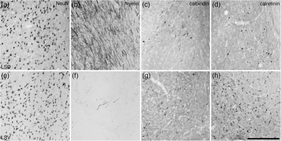
Photomicrographs of coronal sections through the lateral septal nucleus, dorsal part (LSD) (a–d) and the lateral septal nucleus, ventral part (LSV) (e–h), stained for neuronal nuclear marker (NeuN; a and e), myelin (b and f), calbindin (c and g), and calretinin (d and h). Clear variations in staining densities for myelin (b and f), calbindin (c and g), and calretinin (d and h) support the division of these regions of the lateral septal nucleus into the two nuclei. In all images, medial is to the left and dorsal to the top. Scale bar in (h) = 250 μm and applies to all. See list for abbreviations