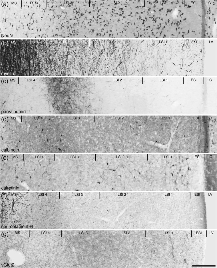
FIGURE 6.

Photomicrographs of coronal sections through the lateral septal nucleus, intermediate part (LSI), showing the four distinct layers (LSI 1–4) in the brain of the tree pangolin stained for neuronal nuclear marker (NeuN; a), myelin (b), parvalbumin (c), calbindin (d), calretinin (e), neurofilament H (f), and vesicular glutamate transporter 2 (vGlut2; g). Using this combination of stains, the four layers could be readily discerned. Note the presence of a cell sparse lamina on the lateral, or external, surface of the septal nuclear complex, which we have termed the external septal lamina (ESl). In all images, medial is to the left and dorsal to the top. Scale bar in (g) = 200 μm and applies to all. See list for abbreviations
