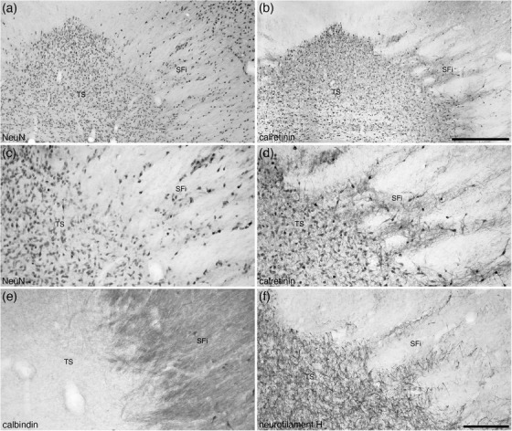
FIGURE 7.

Photomicrographs at lower (a and b) and higher (c–f) magnifications of coronal sections through the triangular septal (TS) and septofimbrial (SFi) nuclei of the tree pangolin stained for neuronal nuclear marker (NeuN; a and c), calretinin (c and d), calbindin (e), and neurofilament H (f). Note the distinct triangular shape of the TS, and while the cells forming the striated appearance of the SFi may be interpreted as extensions of the TS neurons (a, c, and f), the difference in morphology of the calretinin neurons (d) between the two nuclei and the difference in calbindin neuropil staining (e) support the distinction of these two nuclei. In all images, medial is to the left and dorsal to the top. Scale bar in (b) = 500 μm and applies to (a) and (b). Scale bar in (f) = 200 μm and applies to (c)–(f)
