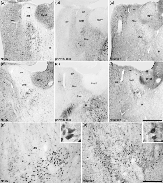
FIGURE 9.

Photomicrographs of the nucleus of the stria medullaris (SM) in coronal sections of the tree pangolin brain stained for neuronal nuclear marker (NeuN; a, d, and g), parvalbumin (b and e), and calretinin (c, f, and h). The low magnification images (a–f) represent adjacent sections with different stains (a–c and d–f, with d–f being 250 μm caudal to a–c) to show the location of the SM in relation to the bed nuclei of the stria terminalis (BNST), thalamic reticular nucleus (R), and dorsal thalamus (DT). At higher magnifications, the SM appears divisible into two parts, a compact (SMc) part located ventrally and a diffuse (SMd) part located dorsally and housing far fewer neurons scattered in the fibers of the stria medullaris thalami (sm). The neurons of these two parts of the SM (g) are calretinin immunopositive (h), with scattered neurons in the more rostral parts being parvalbumin immunopositive (b). Insets (g) and (h) are higher magnification images showing the morphology of the neurons in the SMc stained for NeuN (inset g) and calretinin (inset h). In all photomicrographs, medial is to the left of the image and dorsal to the top. Scale bar in (f) = 1 mm and applies to a–f. Scale bar in (h) = 200 μm and applies to (g) and (f). Scale bar in inset (h) = 20 μm and applies to both insets. See list for abbreviations
