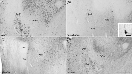
FIGURE 10.

Photomicrographs of coronal sections through the bed nucleus of the anterior commissure (BAC) and the medial para‐tractal region (PtRm) of the bed nuclei of the stria terminalis of the tree pangolin brain stained for neuronal nuclear marker (NeuN; a), parvalbumin (b), calbindin (c), and calretinin (d). The BAC is not composed of a great number of neurons (a), many of which appear to be parvalbumin‐immunopositive (b). PtRm represents a region in the BNST that has a low neuronal density (a, c, and e), but appears to contain many parvalbumin immunopositive fibers (b). In all photomicrographs, medial is to the left and dorsal to the top. Scale bar in (d) = 500 μm and applies to all. Scale bar in inset (b) = 20 μm. See list for abbreviations
