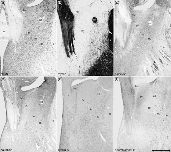
FIGURE 11.

Low magnification photomicrographs of coronal sections through the tree pangolin brain showing the location of the nuclei forming the rostral portion of the bed nuclei of the stria terminalis including the rostral medial (RM), principal (Pr), rostral intermediate (RI), and rostral lateral (RL) divisions stained for neuronal nuclear marker (NeuN; a), myelin (b), calbindin (c), calretinin (d), orexin‐A (e), and neurofilament H (f). Consistency in the variation of the patterns of staining allowed the delineation of these four nuclei, but we note here that the borders between the nuclei are not sharp, with the nuclei appearing to undergo transitions at their borders. In all images, medial is to the left and dorsal to the top. Scale bar in (f) = 1 mm and applies to all. See list for abbreviations
