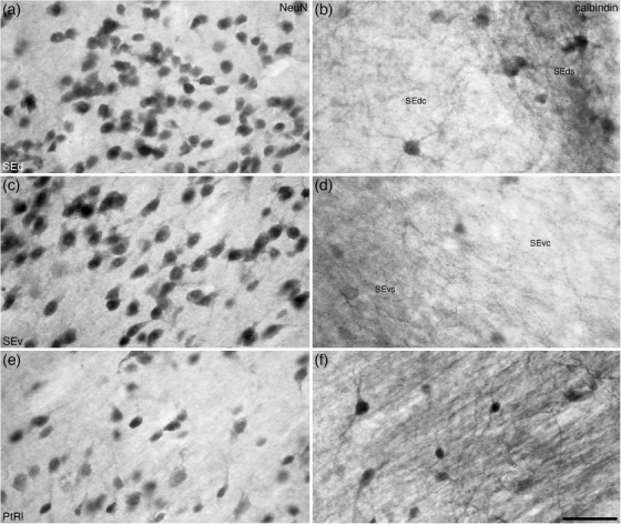
FIGURE 14.

High magnification photomicrographs of coronal sections through the striatal extension (SE) and lateral para‐tractal region (PtRl) of the bed nuclei of the stria terminalis of the tree pangolin brain stained for neuronal nuclear marker (NeuN; a, c, and e) and calbindin (b, d, and f). Note the presence of core (SEdc/SEvc) and surround (SEds/SEvs) subdivisions in both the dorsal (SEd) and ventral (SEv) parts of the SE, despite similar neuronal densities (a and c) in these regions. A greater density of calbindin immunopositive structures are observed in the SEds/SEvs compared with the SEdc/Sevc in both the Sed (b) and Sev (d). The lower neuronal density € and homogenous high density of calbindin immunopositive structures (f) in the PtRl distinguish this region from the SE. In all photomicrographs, medial is to the left and dorsal to the top. Scale bar in (f) = 50 μm and applies to all. See list for abbreviations
