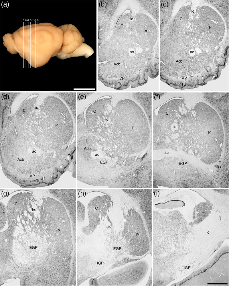
FIGURE 15.

(a) Lateral view of the tree pangolin brain showing the levels at which the coronal sections imaged in (b)–(i) were taken. Scale bar in (a) = 1 cm and applies to (a) only. (b)–(i) A series of rostral to caudal low magnification photomicrographs of neuronal nuclear marker immunostained sections, each section being approximately 1000 μm apart. These images show the location of the components of the ventral striatopallidal complex, including the nucleus accumbens (Acb), the ventral pallidum (VP), and the olfactory tubercule (Tu), as well as the components of the dorsal striatopallidal complex, including the caudate nucleus (C), putamen nucleus (P), external globus pallidus (EGP), and internal globus pallidus (IGP; also referred to as the intrapeduncular or entopeduncular nucleus). Note that while the internal capsule (ic) is present, this pathway is only fully consolidated at the posterior aspects of the dorsal striatopallidal complex. In all photomicrographs, medial is to the left and dorsal to the top. Scale bar in (i) = 2 mm and applies to (b)–(i). See list for abbreviations
