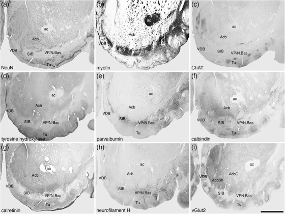
FIGURE 16.

Low magnification photomicrographs showing the location of the nucleus accumbens (Acb), ventral pallidum (VP)/nucleus basalis (N.Bas), substantia innominata, basal part (SIB), vertical diagonal band (VDB), and olfactory tubercle (Tu) in coronal sections of the tree pangolin brain stained for neuronal nuclear marker (NeuN; a), myelin (b), choline acetyltransferase (ChAT; c), tyrosine hydroxylase (d), parvalbumin (e), calbindin (f), calretinin (g), neurofilament H (h), and vesicular glutamate transporter 2 (vGlut2; i), where the core (AcbC) and shell (AcbSh) are evident. Note that the VP/N.Bas and SIB are in a region of high myelin density (b) as well as a high density of parvalbumin (e) and neurofilament H (h) immunoreactive structures. The overlap of the cholinergic neurons of the N.Bas (c) with parvalbumin and neurofilament H immunoreactive structures has been used to delineate the VP, whereas the absence of cholinergic neurons is used to delineate the SIB. In all images, medial is to the left and dorsal to the top. Scale bar in (i) = 2 mm and applies to all. See list for abbreviations
