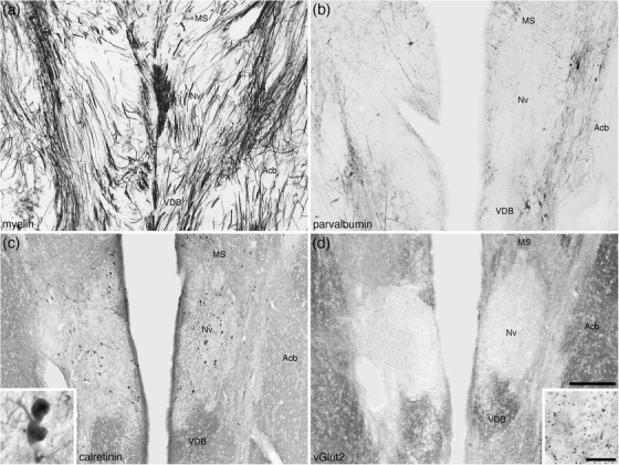
FIGURE 21.

Low (a–d) and higher (insets c and d) magnification photomicrographs of coronal sections through the navicular nucleus of the basal forebrain (Nv) of the tree pangolin brain stained for myelin (a), parvalbumin (b), calretinin (c), and vesicular glutamate transporter 2 (vGlut2; d). Note the pale neuropil staining for parvalbumin (b), calretinin (c), and vGlut2 (d) that delineate this nucleus from the medial septal nucleus (MS) dorsally, the vertical diagonal band (VDB) ventrally and the nucleus accumbens (Acb) laterally. A low density of calretinin immunopositive neurons are observed in the Nv (c) as well as a low density of vGlut2 immunopositive boutons (d). In all photomicrographs, medial is at the center of the image and dorsal to the top. Scale bar in (d) = 250 μm and applies to all. Scale bar in inset (d) = 20 μm and applies to both insets. See list for abbreviations
