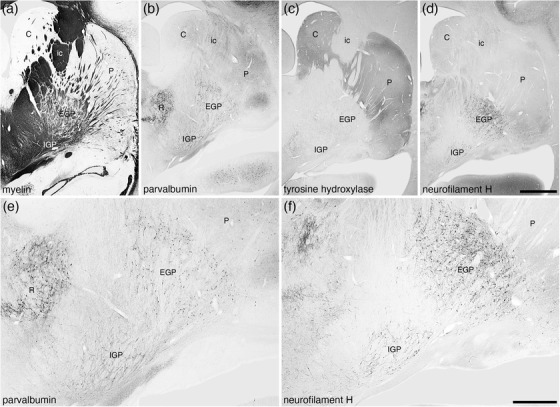
FIGURE 24.

Low magnification photomicrographs of coronal sections through the dorsal striatopallidal complex of the tree pangolin brain stained for myelin (a), parvalbumin (b and e), tyrosine hydroxylase (c), and neurofilament H (d and f). These images demonstrate the location of the components of this complex including the caudate nucleus (C), putamen nucleus (P), external globus pallidus (EGP), internal globus pallidus (IGP), and the internal capsule (ic). Panels (e) and (f) show higher magnification images of the EGP and IGP stained for parvalbumin (e) and neurofilament H (f), which clearly demonstrate these nuclei. In all photomicrographs medial is to the left and dorsal to the top. Scale bar in (d) = 2 mm and applies to (a)–(d). Scale bar in (f) = 1 mm and applies to (e) and (f). See list for abbreviations
