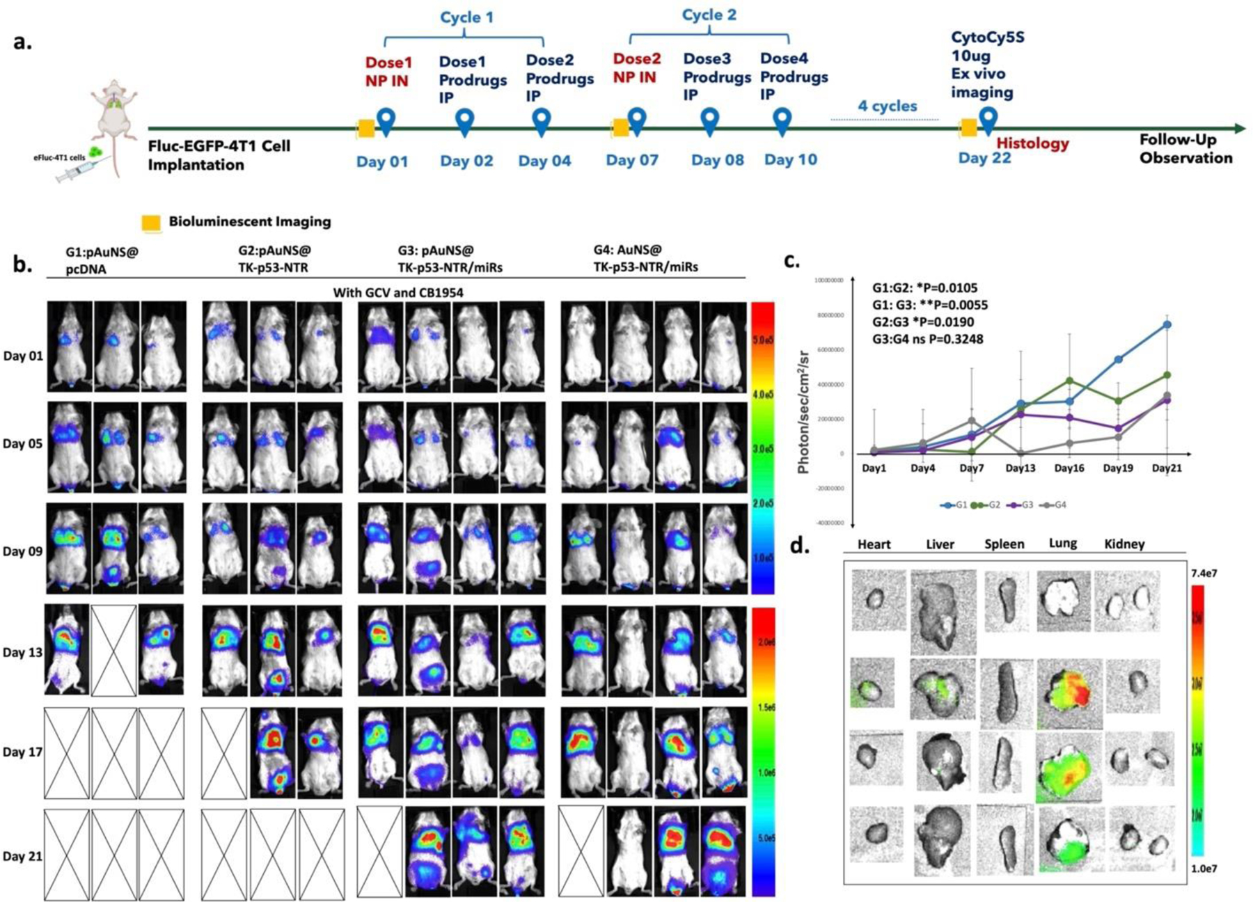
Figure 5. In vivo estimation of the therapeutic potential of the TK-p53-NTR gene combined with therapeutic miRs in the presence of GCV/CB1954 prodrugs in mice bearing TNBC lung metastatic xenografts and delivered using uPA-targeted AuNS NPs.

(a) Schematic outline of the in vivo study design for imaging and treatment timelines. (b) Bioluminescence imaging (BLI) of Balb/c mice bearing 4T1 TNBC lung metastasis xenografts expressing Fluc-eGFP reporter gene at multiple time points (G1 denotes pcDNA delivered by pAuNS NPs, G2 denotes TK-p53-NTR gene alone delivered by pAuNS NPs, G3 denotes TK-p53-NTR and miRs (antimiR-21, antimiR-10b and miR-100) delivered by pAuNS NPs, and G4 denotes TK-p53-NTR gene and miRs delivered using non-targeted AuNS NPs. All mice received a combination of prodrugs (GCV and CB1954). (c) Tumor volume estimation by the quantitative plot of the BLI intensity during the treatments. (d) TK-p53-NTR gene delivery evaluation in mice by ex vivo CytoCy5S fluorescence imaging at the termination of treatment (n=3–5/group; data are presented as mean ± SD; the significance of comparisons, as indicated, is drawn using One-way ANOVA with Bonferroni post hoc test. Adjusted p - values were considered statistically significant if p- values were < 0.05. The symbols indicating statistical significance are as follows: ns- represents non-significant difference, * represents p< 0.05, ** represents p< 0.01, *** represents p< 0.001, and **** represents p< 0.0001).
