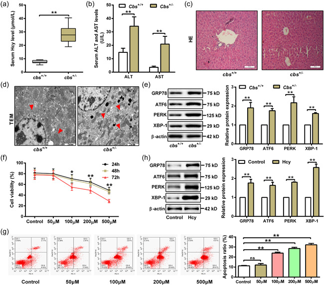
Figure 1.

Homocysteine (Hcy) promotes endoplasmic reticulum (ER) stress and apoptosis of hepatocytes leading to liver injury. (a and b) Serum levels of Hcy, aspartate aminotransferase (AST) and alanine transaminase (ALT) in cbs +/+ and cbs +/ − mice fed with high‐methionine diet were measured by automatic biochemical analyzer (n = 6). (c) Liver injury was evaluated by hematoxylin and eosin (H&E) staining of liver slices from cbs +/+ and cbs +/ − mice. Scale bar, 100 μM. (d) Transmission electron microscopy (TEM) was performed to observe endoplasmic reticulum in liver slices from cbs +/+ and cbs +/‐ mice. Red arrows indicate hyperplasia of rough endoplasmic reticulum in hepatocytes. Scale bar, 2 μM. (e) Western blot was used to measure GRP78, ATF6, PERK, and XBP‐1 protein expression in liver tissue of cbs +/+ and cbs +/ − mice (n = 6). (f) Cell viability was detected by MTT assay in hepatocytes treated with Hcy at indicated concentrations (0, 50, 100, 200, and 500 μM) for 24, 48, and 72 h (n = 3). (g) Apoptosis rate was measured by flow cytometry after treating hepatocytes with different concentration of Hcy (0, 50, 100, 200, and 500 μM) for 48 h (n = 3). (h) The protein expression of GRP78, ATF6, PERK, and XBP‐1 were detected by western blot after treating hepatocytes with 100 μM Hcy for 48 h (n = 3). Data represents mean ± SD, **p <0 .01. MTT, 3‐(4,5‐dimethyl‐2‐thiazolyl)‐2,5‐diphenyl‐2H‐tetrazolium bromide; SD, standard deviation
