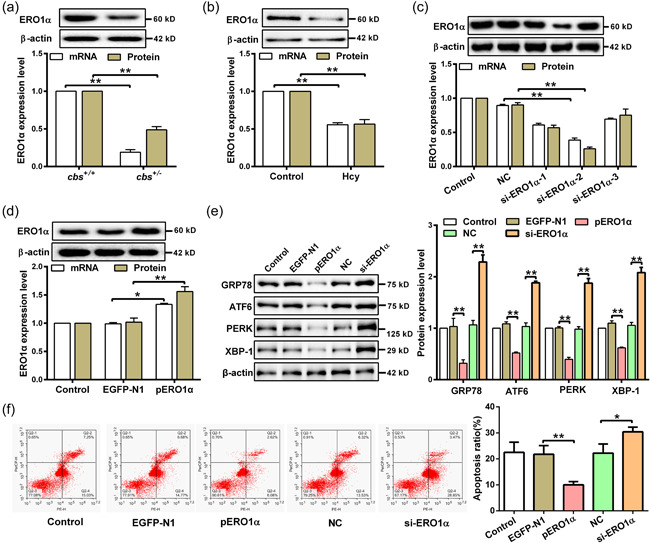
Figure 2.

Hcy induces ER stress and apoptosis by downregulation of ERO1α in hepatocyte. (a and b) Western blot and qRT‐PCR were used to detect the mRNA and protein expression of ERO1α in the liver tissue of cbs +/+ and cbs +/ − mice (n = 6) and hepatocytes treated with 100 μM Hcy (n = 3). (c) The expression of ERO1α was examined by qRT‐PCR and western blot in hepatocytes transfected with siRNAs targeting ERO1α (si‐ERO1α‐1/2/3) (n = 3). (d) ERO1α expression in hepatocytes transfected with ERO1α‐overexpressed plasmids (pERO1α) or negative control vector (EGFP‐N1) was detected by qRT‐PCR and western blot (n = 3). (e) The protein expression of GRP78, ATF6, PERK, and XBP‐1 were measured by western blot in hepatocytes transfected with pERO1α or si‐ERO1α in the presence of Hcy (n = 3). (f) Flow cytometry was used to detect the apoptosis rate of hepatocytes transfected with pERO1α or si‐ERO1α in the presence of Hcy (n = 3). Data represents mean ± SD, *p < 0.05, **p < 0 .01. ER, endoplasmic reticulum; ERO1α, endoplasmic reticulum oxidoreductase 1α; Hcy, homocysteine; qRT‐PCR, quantitative real‐time polymerase chain reaction; SD, standard deviation
