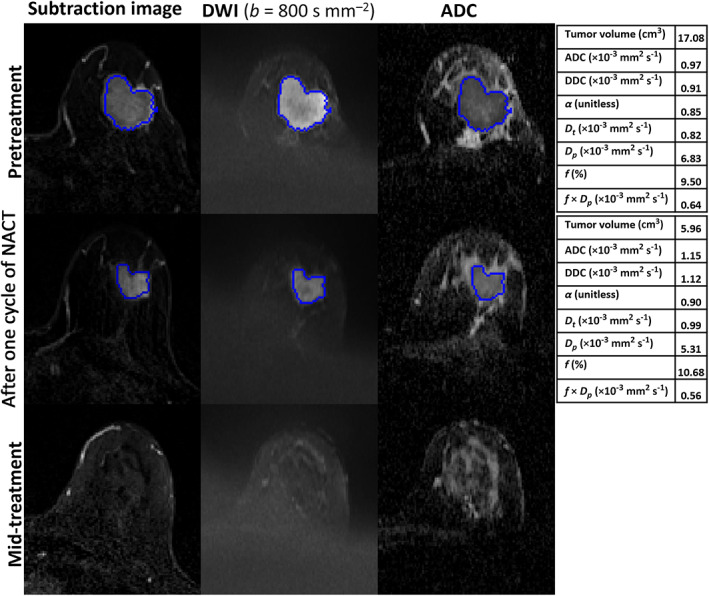
FIGURE 3.

MRI scans of a 45‐year‐old woman with invasive ductal carcinoma in the left breast who showed a complete pathological response after surgery (residual cancer burden [RCB]‐0). Each row includes images acquired at pretreatment, after one cycle of neoadjuvant chemotherapy (NACT), and at mid‐treatment. The seeded region of interest (ROI) for the given slice is shown in blue. The tables represent the parameter estimates of monoexponential, stretched‐exponential model (SEM) and intravoxel incoherent motion (IVIM) model at each time‐point. At mid‐treatment, no tumor was visible on the dynamic contrast‐enhanced (DCE) and diffusion‐weighted (DW) images obtained.
