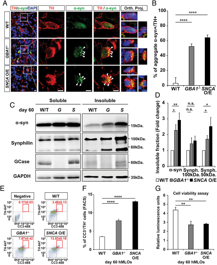
FIGURE 3.

GBA1 −/− and SNCA overexpressing (O/E) human midbrain‐like organoids (hMLOs) show α‐synuclein (α‐syn) aggregation and apoptosis‐driven neurodegeneration. (A) Cryosections of wild‐type (W/T), GBA1 −/−, and SNCA O/E hMLOs at day 60 stained with specific antibodies against tyrosine hydroxylase (TH) and α‐syn. The merged images show orthogonal Z‐stack projections (Orth. Proj.). Scale bars = 10μm. (B) Percentage of TH+ cells that have aggregated α‐syn (mean ± standard error of the mean [SEM]; ****p < 0.0001; n = 3). (C) Western blots of detergent‐soluble and detergent‐insoluble α‐syn, synphilin‐1, and glucocerebrosidase (GCase) proteins in wild‐type, GBA1 −/− (G), and SNCA O/E (S) hMLOs. Glyceraldehyde‐3‐phosphate dehydrogenase (GAPDH) was used as a loading control. (D) Quantification of changes in the compositions of insoluble protein fractions (mean ± SEM; n.s. = no significance, *p < 0.05, **p < 0.005; n = 4). (E) Fluorescence‐activated cell sorting (FACS) analysis of cells dissociated from day 60 hMLOs to quantify the percentages of CC3+/TH+ cells (mean ± SEM; n = 3). A threshold gate was drawn based on a secondary antibody‐only negative control. (F) Quantification of CC3+/TH+ cells by FACS revealed a significant increase in apoptotic midbrain dopaminergic neurons in PD‐related hMLOs compared to non–Parkinson disease–related hMLOs (mean ± SEM; ****p < 0.0001; n = 3). (G) Cell viability assay for hMLOs (mean ± SEM; **p = 0.005, 0.004; n = 5). DAPI = 4,6‐diamidino‐2‐phenylindole. [Color figure can be viewed at www.annalsofneurology.org]
