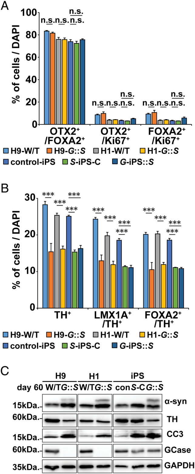
FIGURE 6.

Characterization of H9‐GBA1 −/−::SNCA overexpressing (O/E) and H1‐GBA1 −/−::SNCA O/E human midbrain‐like organoids (hMLOs), SNCA triplication‐induced pluripotent stem cell (iPSC) hMLOs with conduritol‐b‐epoxide (CBE) treatment (S‐iPS‐C), and GBA1‐iPSC hMLOs with SNCA overexpression (G‐iPS::S). (A) Quantification of cells expressing combinations of the midbrain progenitor markers OTX2, FOXA2, and Ki67 in day 60 H9‐ and H1‐GBA1 −/−::SNCA O/E hMLOs (G::S), SNCA triplication iPSC‐derived hMLOs with or without CBE treatment, and GBA1 mutant iPSC‐derived hMLOs (mean ± standard error of the mean [SEM]; n.s. = no significance; n = 3). (B) Quantification of cells expressing combinations of the midbrain progenitor markers FOXA2 and LMX1A with the dopaminergic neuron marker tyrosine hydroxylase (TH) in day 60 H9‐ and H1‐GBA1 −/−::SNCA O/E hMLOs, SNCA triplication iPSC‐derived hMLOs with or without CBE treatment, and GBA1 mutant iPSC‐derived hMLOs (mean ± SEM; ***p < 0.001; n = 3). (C) Western blots for α‐synuclein (α‐syn), TH, CC3, and glucocerebrosidase (GCase) in hMLOs derived from H9‐ and H1‐GBA1 −/−::SNCA O/E hMLOs, SNCA triplication iPSC‐derived hMLOs with or without CBE treatment, and GBA1 mutant iPSC‐derived hMLOs. DAPI = 4,6‐diamidino‐2‐phenylindole; GAPDH = glyceraldehyde‐3‐phosphate dehydrogenase; W/T = wild type. [Color figure can be viewed at www.annalsofneurology.org]
