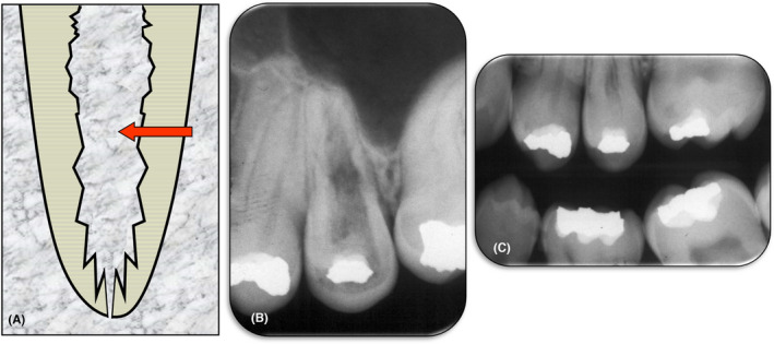
FIGURE 4.

Internal Replacement Resorption. (A) Schematic diagram; (B) Periapical radiograph of tooth 25 with extensive internal replacement resorption throughout the entire tooth; (C) A bitewing radiograph taken 3 years earlier showing the pulp chamber and coronal portion of the root of the same tooth. The irregular shape and appearance of the pulp space suggests that the internal replacement resorption had likely already commenced. A comparison of these two radiographs indicates that over the 3‐year period, the resorptive process continued and bone‐like tissue replaced the lost dentin throughout the length of the root and extending into the crown of the tooth
