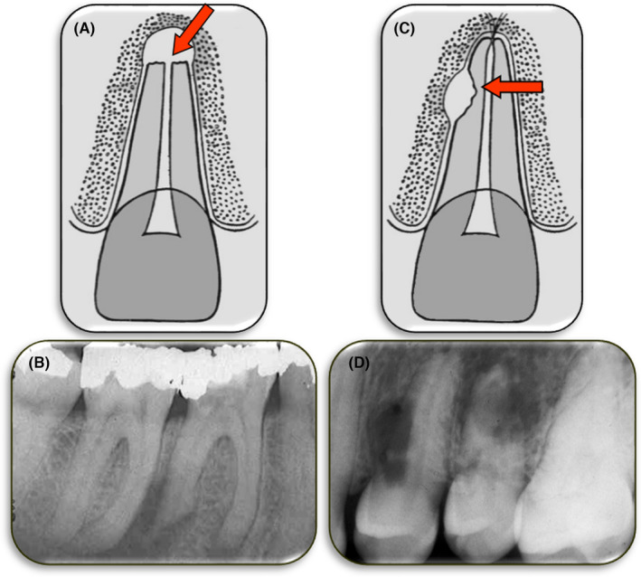
FIGURE 7.

Two types of External Inflammatory Resorption. (A) Diagram demonstrating external apical inflammatory resorption; (B) Periapical radiograph of tooth 36 with extensive external apical inflammatory resorption of both the mesial and distal roots; (C) Diagram demonstrating external lateral inflammatory resorption; (D) Radiograph of teeth 24 and 25 with extensive external apical inflammatory resorption of their roots
