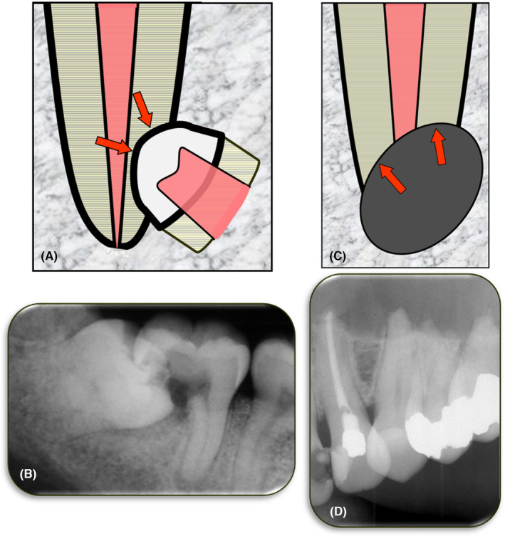
FIGURE 12.

External Pressure Resorption. (A) Schematic diagram of external pressure resorption caused by an impacted tooth located adjacent to the tooth undergoing resorption; (B) Periapical radiograph of tooth 47 with extensive pressure resorption caused by the mesially‐angled impacted tooth 48; (C) Schematic diagram of external pressure resorption caused by a tumor, cyst or other pathological condition located adjacent to the tooth undergoing resorption; (D) Periapical radiograph showing pressure resorption of the apices of teeth 23, 24, 25 and 26 which was caused by an odontogenic keratocyst in the left maxilla
