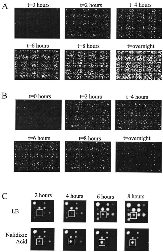
FIG. 1.
Images of duplicate LuxArray 1.0 cellular bioluminescent reporter arrays. Following the spotting of the E. coli strains containing reporter gene fusions onto membranes on LB agar plates and growth for 6 h, the membranes were moved to LB medium plates (A) or LB medium plates containing 5 μg of nalidixic acid/ml (B). The images were taken as described in Materials and Methods immediately after moving the membrane and subsequently at 2, 4, 6, and 8 h and after overnight incubation. A magnification of the 16 spots in the D-4 primary location from panels A and B is shown in panel C. The spot at the secondary location of row 3, column 2, containing cells with upregulated gene fusion dinB-luxCDABE is boxed.

