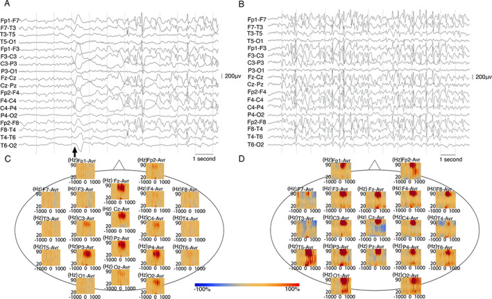
FIGURE 1.

(A and B) A 7 year‐old male with NS (participant s3) onset at age 6 years. (A) Ictal EEG shows generalized slow wave followed by electrodecrement. EEG background between each head nod is marked by generalized spike and wave discharges. (B) Interictal EEG shows 2‐ 3 Hz generalized spike and wave discharges. Both EEGs are shown in bipolar montage. Low frequency filter: 0.053 Hz, high frequency filter: 70 Hz Black arrows indicate head drops. C and D: Time frequency analysis of ictal head nod showed augmentation of gamma activity on scalp EEG of children with NS from South Sudan and Tanzania. (C) In an 18 year‐old male with NS (participant s8) from South Sudan, time‐frequency plot derived from 70 head nodding episodes showed maximal augmentation of gamma (30 Hz‐70 Hz) at the centro‐parietal electrodes during the head drops. (D) In a 12 year‐old male (participant s4) from Tanzania, time‐frequency plot derived from 17 head nodding episodes showing augmentation of gamma‐activity. Blue color indicates attenuation of amplitude and red color indicates augmentation of amplitude in the corresponding time‐frequency bin relative to the baseline.
