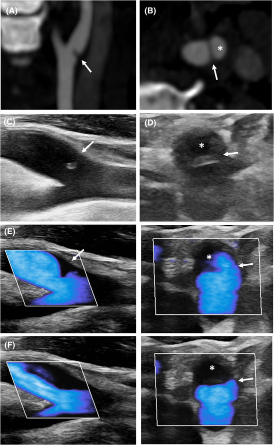
FIGURE 1.

Typical appearance of carotid web. Longitudinal (A) and axial (B) views of carotid web (CaW) on CT‐angiography. The CaW (arrow) delimits a nest (*). (C) Longitudinal view on B‐mode shows an isoechoic lesion protruding into the arterial lumen (arrow). (D) Corresponding axial view on B‐mode reveals a septum (arrow) delimiting a nidus (*). (E) Microflow imaging delineates the CaW during systole (E) with downstream low flow velocities during diastole (F)
