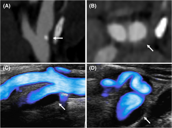
FIGURE 5.

Atherosclerotic plaque on microflow imaging (MFI). Atherosclerotic plaque on CTA (A and B) and MFI (C and D). The MFI‐mode during systole delineates a mostly anechoic plaque with a small calcification and without area of low flow on longitudinal view (C). The axial incidence shows a convex anechoic thickening of the artrial wall without nidus (D)
