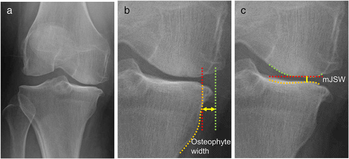
FIGURE 1.

Measurement methods used with radiographs. (a) Representative radiograph—anteroposterior view of a subject standing with the knee extended. (b) Measurement of osteophyte width. A line along the medial edge of the tibia (orange line) is drawn. A vertical line (red line) is then drawn from the point of the intersection of that line and the articular surface. A third line (green line) is drawn from the inner edge of the osteophyte of the medial tibia. The distance between the red and green lines is defined as the osteophyte width (yellow arrow). (c) Measurement of the mJSW. A bright radio‐dense zone (orange line), defined at the anterior edge of the medial tibia, was drawn. A horizontal straight line (red line) was also drawn at the lowest end of the medial femoral condyle (green line). The minimum distance between these two lines was defined as mJSW (yellow line). mJSW, minimum joint space width at the medial compartment.
