
FIGURE 2.
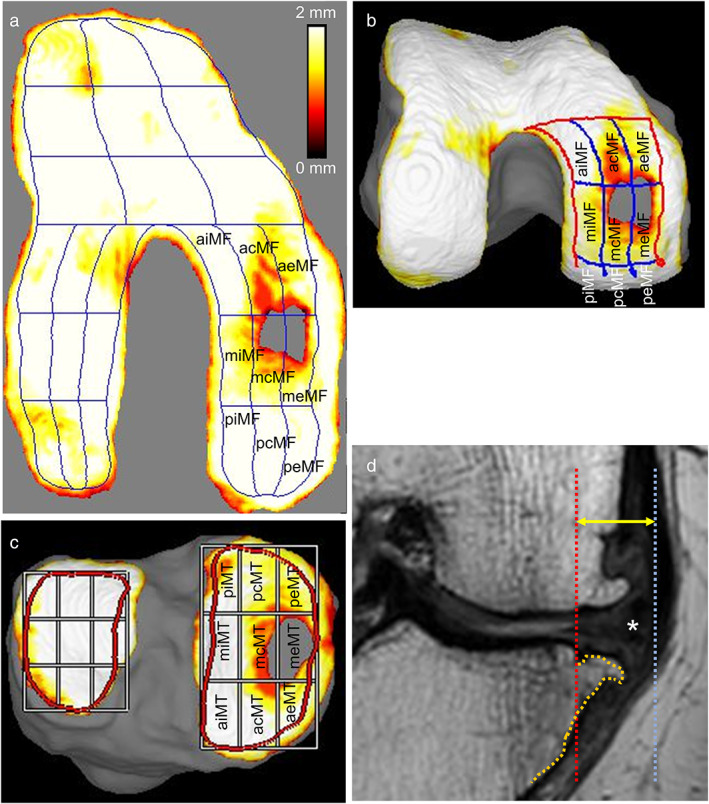
Subregions and MME measurement methods in MRI. The knee shown in Fig. 1 is further examined. (a) Radially projected cartilage thickness mapping of the femoral cartilage and subregions of the medial femoral cartilage. The MF region is the combined area of the nine MF subregions. Cartilage thickness is shown with a color bar. (b) Three‐dimensional femoral cartilage viewed from below along the long axis of the femur. Subregions of the medial femoral cartilage are also shown. (c) Three‐dimensional tibial cartilage and subregions of the medial tibial cartilage. The MT region is the combined area of the nine MT subregions. (d) Measurement of MME. A line along the medial edge of the tibia (orange line) is drawn using a coronal plane MR. A vertical line (red line) is then drawn from the point of the intersection of that line, excluding the osteophytes, and the articular surface. A third line (blue line) is drawn from the outer edge of the MM (asterisk). The distance between the red and blue lines is defined as the MME distance (yellow arrow). acMF, anterior central MF; acMT, anterior central MT; aeMF, anterior external MF; aeMT, anterior external MT; aiMF, anterior internal MF; aiMT, anterior internal MT; mcMF, middle central MF; mcMT, middle central MT; meMF, middle external MF; meMT, middle external MT; MF, medial femoral; miMF, middle internal MF; miMT, middle internal MT; MME, medial meniscus extrusion; MRI, magnetic resonance imaging; MT, medial tibial; pcMF, posterior central MF; pcMT, posterior central MT; peMF, posterior external MF; peMT, posterior external MT; piMF, posterior internal MF; piMT, posterior internal MT.