Fig. 6.

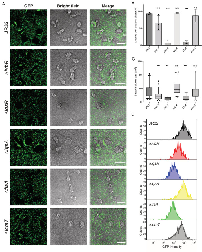
LvbR, LqsR and FlaA determine L. pneumophila cluster formation on A. castellanii. Biofilms of L. pneumophila JR32, ∆lvbR, ∆lqsR, ∆lqsA, ∆flaA or ∆icmT mutant strains harbouring pNT28 (constitutive production of GFP) were grown for 6 days in AYE medium at 25°C, and A. castellanii was added to preformed biofilms.
A. Bacterial adherence and cluster formation were monitored in biofilms containing amoebae by confocal microscopy above the dish bottom. The images shown are representative of three independent experiments. Scale bars, 30 μm.
B. Percentage of amoebae with bacterial cluster (or cluster‐positive amoebae) in the total amoebae population. The columns display the mean value of three biological replicates (in dots) with standard deviations.
C. Quantification by fluorescence microscopy of cluster size on cluster‐positive amoebae. The box‐and‐whisker plots display 5th to 95th percentiles (whiskers), median and quartiles (box) of the pooled results from three biologically independent experiments with a total analysed number of amoebae n = 124 (JR32), n = 60 (∆lvbR), n = 9 (∆lqsR), n = 66 (∆lqsA), n = 8 (∆flaA), or n = 48 (∆icmT). For (B) and (C): n.s., not significant; *p ≤ 0.05; **p ≤ 0.01; ***p ≤ 0.001; one‐way ANOVA with Tukey post‐test; JR32 vs. mutant strains.
D. Quantification by flow cytometry (counts vs. fluorescence intensity) of amoeba‐associated, GFP‐positive L. pneumophila (JR32, ∆lvbR, ∆lqsR, ∆lqsA, ∆flaA or ∆icmT harbouring pNT28) using FlowJo software (protocol #1, see Materials and Methods).
