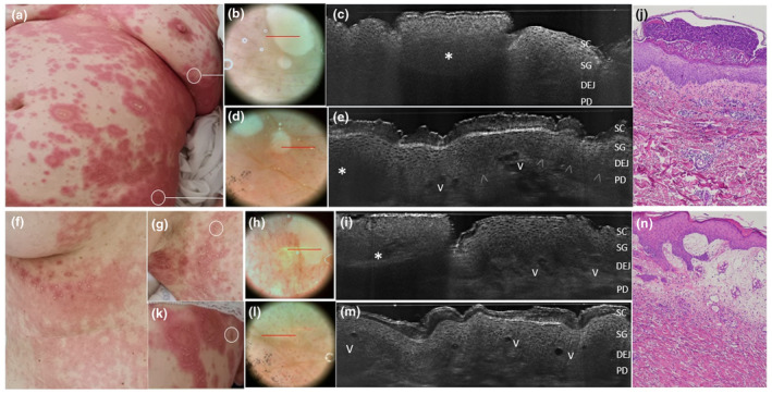
Figure 2.

Generalized pustular figurate erythema in women aged 57 years (a‐e) and 50 years (f‐n) triggered by hydroxychloroquine. Clinical examination reveal annular to polycyclic erythematous‐oedematous plaques distributed on the trunk, proximal extremities and flexural areas in both cases (a, f, g). Under in vivo 2D LC‐OCT examination performed at lesional margins form both patients (c,i), pustules appeared as intraepidermal (c) or subcorneal (i) areas of roundish shape, well‐defined borders and hyper‐reflective homogenous content due to a dense neutrophils collection (asterisks) generating a posterior shadow. Dilated vessels are visible as black a‐reflective areas (V) in perilesional skin (k) along with papillary dermal vessels and spongiosis (e, m). Matched histology showing a subcorneal pustule (l, haematoxylin‐eosin, OM 40×) and a perilesional skin with edematous papillary dermis with ectatic vessels and inflammatory cells (n, haematoxylin‐eosin, OM 100×). Papillary vessels correspond to the red dots seen in dermoscopy (e, m) [SC, stratum corneum; SG, stratum granulosum; DEJ, dermal‐epidermal junction; PD, papillary dermis; V, capillary vessels; arrowheads: DEJ profile. The red line inside polarized dermoscopy images 15× (b/e/i/o, box) corresponds to the LC‐OCT vertical frame].
