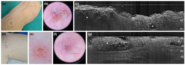
Figure 4.

Clinical, dermoscopic and LC‐OCT appearance of Intraepidermal IgA pustulosis (IAD) lesions in two women, one aged 61 years with the Subcorneal Pustular Dermatosis‐like variant (IAD‐SPD) (a‐c) and one aged 42 years with the Intraepidermal neutrophilic IgA dermatosis‐like variant (IAD‐IEN) (d‐g). In vivo 2D LC‐OCT reveals two upper‐epidermal spongiotic‐multilocular pustules with ill‐defined borders (c, asterisks) in IAD‐SPD, while intraepidermal unilocular pustules with well‐defined borders (g, asterisks) in IAD‐IEN variant. [SC, stratum corneum; SG, stratum granulosum; DEJ, dermal‐epidermal junction; arrowheads: DEJ profile; PD, papillary dermis. The red line inside polarized dermoscopy 15× (b, f) corresponds to the LC‐OCT vertical frame].
